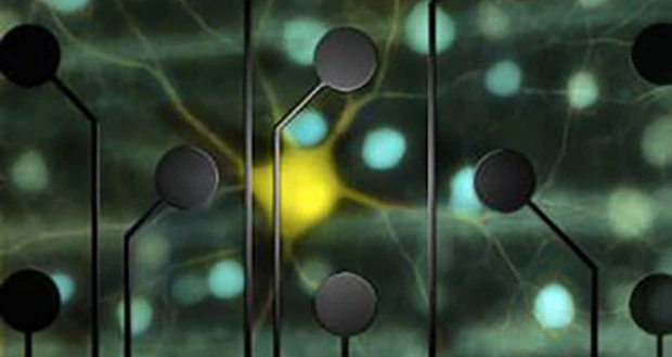
Bethesda, Maryland – In laboratory tests, researchers have used electrical stimulation of retinal cells to produce the same patterns of activity that occur when the retina sees a moving object. Although more work remains, this is a step toward restoring natural, high-fidelity vision to blind people, the researchers say. The work was funded in part by the National Institutes of Health.
Just 20 years ago, bionic vision was more a science fiction cliché than a realistic medical goal. But in the past few years, the first artificial vision technology has come on the market in the United States and Western Europe, allowing people who’ve been blinded by retinitis pigmentosa to regain some of their sight. While remarkable, the technology has its limits. It has enabled people to navigate through a door and even read headline-sized letters, but not to drive, jog down the street, or see a loved one’s face.
A team based at Stanford University in California is working to improve the technology by targeting specific cells in the retina – the neural tissue at the back of the eye that converts light into electrical activity.
“We’ve found that we can reproduce natural patterns of activity in the retina with exquisite precision,” said E.J. Chichilnisky, Ph.D., a professor of neurosurgery at Stanford’s School of Medicine and Hansen Experimental Physics Laboratory. The study was published in Neuron, and was funded in part by NIH’s National Eye Institute (NEI) and National Institute of Biomedical Imaging and Bioengineering (NIBIB).
The retina contains several cell layers. The first layer contains photoreceptor cells, which detect light and convert it into electrical signals. Retinitis pigmentosa and several other blinding diseases are caused by a loss of these cells. The strategy behind many bionic retinas, or retinal prosthetics, is to bypass the need for photoreceptors and stimulate the retinal ganglion cell layer, the last stop in the retina before visual signals are sent to the brain.
Several types of retinal prostheses are under development. The Argus II, which was developed by Second Sight Therapeutics with more than $25 million in support from NEI, is the best known of these devices. In the United States, it was approved for treating retinitis pigmentosa in 2013, and it’s now available at a limited number of medical centers throughout the country. It consists of a camera, mounted on a pair of goggles, which transmits wireless signals to a grid of electrodes implanted on the retina. The electrodes stimulate retinal ganglion cells and give the person a rough sense of what the camera sees, including changes in light and contrast, edges, and rough shapes.
“It’s very exciting for someone who may not have seen anything for 20-30 years. It’s a big deal. On the other hand, it’s a long way from natural vision,” said Dr. Chichilnisky, who was not involved in development of the Argus II.
Current technology does not have enough specificity or precision to reproduce natural vision, he said. Although much of visual processing occurs within the brain, some processing is accomplished by retinal ganglion cells. There are 1 to 1.5 million retinal ganglion cells inside the retina, in at least 20 varieties. Natural vision – including the ability to see details in shape, color, depth and motion – requires activating the right cells at the right time.
The new study shows that patterned electrical stimulation can do just that in isolated retinal tissue. The lead author was Lauren Jepson, Ph.D., who was a postdoctoral fellow in Dr. Chichilnisky’s former lab at the Salk Institute in La Jolla, California. The pair collaborated with researchers at the University of California, San Diego, the Santa Cruz Institute for Particle Physics, and the AGH University of Science and Technology in Krakow, Poland.
They focused their efforts on a type of retinal ganglion cell called parasol cells. These cells are known to be important for detecting movement, and its direction and speed, within a visual scene. When a moving object passes through visual space, the cells are activated in waves across the retina.
The researchers placed patches of retina on a 61-electrode grid. Then they sent out pulses at each of the electrodes and listened for cells to respond, almost like sonar. This enabled them to identify parasol cells, which have distinct responses from other retinal ganglion cells. It also established the amount of stimulation required to activate each of the cells. Next, the researchers recorded the cells’ responses to a simple moving image – a white bar passing over a gray background. Finally, they electrically stimulated the cells in this same pattern, at the required strengths. They were able to reproduce the same waves of parasol cell activity that they observed with the moving image.
“There is a long way to go between these results and making a device that produces meaningful, patterned activity over a large region of the retina in a human patient,” Dr. Chichilnisky said. “But if we can handle the many technical hurdles ahead, we may be able to speak to the nervous system in its own language, and precisely reproduce its normal function.”
Such advances could help make artificial vision more natural, and could be applied to other types of prosthetic devices, too, such as those being studied to help paralyzed individuals regain movement. NEI supports many other projects geared toward retinal prosthetics.
“Retinal prosthetics hold great promise, but this research is a marathon, not a sprint,” said Thomas Greenwell, Ph.D., a program director in retinal neuroscience at NEI. “This important study helps illustrate the challenges of restoring high-quality vision, one group’s progress toward that goal, and the continued need to for the entire field to keep innovating.”
The study was funded by NIH grants EY012171 and EB004410, the National Science Foundation, the McKnight Foundation, the San Diego Foundation Blasker Award, and the Polish government.
Reference
Jepson LH et al. “High-Fidelity Reproduction of Spatiotemporal Visual Signals for Retinal Prosthesis.” Neuron, June 2014. DOI: 10.1016/j.neuron.2014.04.044
Latest from Today's Medical Developments
- Mazak’s new Customer Solutions Center
- maxon's High Efficiency Joint (HEJ) Drive
- BGS Beta-Gamma-Service installs E-Beam accelerator in US facility
- Trelleborg Medical Solutions advanced solutions for medtech
- MICRO celebrates 80 years of advanced manufacturing for every generation
- Restoring the competitive edge with innovations
- Look into the future of manufacturing in roundtable webinar
- Advancing R&D of fully automated insulin delivery systems


